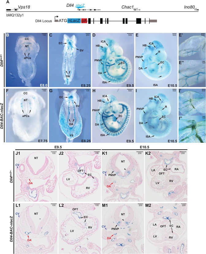
Fig. 1.
Comparative Dll4 expression during early embryonic development. (A) Schematic of the BAC transgene used for generating the Dll4-BAC-nlacZ mouse line, with a magnified schematic of the nuclear LacZ insertion at the ATG start site of Dll4. (B-E) β-gal activity in E7.75-E10.5 Dll4lacZ/+ mouse embryos, ventral (B,C) and sagittal (D,E) views. (F-I) β-gal activity in E7.75-E10.5 Dll4-BAC-nlacZ mouse embryos, ventral (F,G) and sagittal (H,I) views. E′ and I′ show β-gal activity in the embryonic yolk sac. E″ and I″ are magnified views of a representative region shown in corresponding panels E′ and I′, respectively. (J-M) Coronal view of X-gal-stained and Eosin-counterstained sections of E9.5 and E10.5 Dll4lacZ/+ and Dll4-BAC-nlacZ embryos. aPCs, aortic progenitor cells; CC, cardiac crescent; CV, cardinal vein; DA, dorsal aorta; EC, endocardium; HB, hindbrain; ICA, internal carotid artery; ISA, intersegmental artery; LA, left atrium; LV, left ventricle; NT, neural tube; OFT, outflow tract; PNVP, perineural vascular plexus; RA, right atrium; RV, right ventricle; SV, sinus venosus; black caret, ventral V2 interneuron population. Units depicted are in μm.

