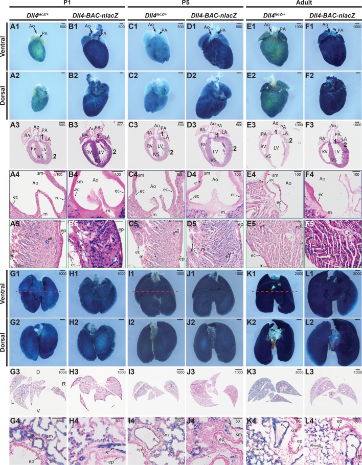
Fig. 7.
Comparative Dll4 expression in postnatal and adult hearts and lungs. (A1-B5) β-gal activity in P1 hearts from (A) Dll4lacZ/+ or (B) Dll4-BAC-nlacZ mice. A1-A2 and B1-B2 show representative wholemount hearts from Dll4lacZ/+ and Dll4-BAC-nlacZ mice, respectively, from ventral and dorsal views. A3 and B3 show β-gal activity in a representative cross-section through the heart, which is magnified accordingly in panels A4-A5 and B4-B5, with activity evident within the endocardial lining of the aorta in both lines (A4,B4), as well as the endocardium, coronary vasculature, and myocardium (A5,B5). (C1-D5) β-gal activity in P5 hearts from (C) Dll4lacZ/+ or (D) Dll4-BAC-nlacZ mice. C1-C2 and D1-D2 show representative wholemount hearts from Dll4lacZ/+ and Dll4-BAC-nlacZ mice, respectively, from ventral and dorsal views. C3 and D3 show β-gal activity in a representative cross-section through the heart, which is magnified accordingly in panels C4-C5 and D4-D5, with signal evident within the endocardial lining of the aorta in both lines, and persisting in the endocardium of the aorta (C4,D4) and chambers, as well as the myocardium and coronary vasculature (C5,D5). (E1-F5) β-gal activity in adult hearts from (E) Dll4lacZ/+ or (F) Dll4-BAC-nlacZ mice. E1-E2 and F1-F2 show representative wholemount hearts from adult Dll4lacZ/+ and Dll4-BAC-nlacZ mice, respectively, from ventral and dorsal views. E3 and F3 show β-gal activity in a representative cross-section through the heart, magnified in panels E4-E5 and F4-F5. β-gal activity is localized to the endocardium of the aortic root Dll4lacZ/+ but absent from Dll4-BAC-nlacZ mice (E4,F4), and present in both lines within the chamber endocardium and coronary vasculature (E5,F5) (asterisks), and within the myocardium. Ao, aorta; ec, endocardium; ep, epicardium; IVS, interventricular septum; LA, left atrium; LV, left ventricle; m, myocardium; PA, pulmonary artery; RA, right atrium; RV, right ventricle; sm, smooth muscle; asterisks – denote lumenized vasculature. (G1-H4) β-gal activity in P1 postnatal lungs from (G) Dll4lacZ/+ or (H) Dll4-BAC-nlacZ mice. G1-G2 and H1-H2 show representative wholemount lungs from Dll4lacZ/+ and Dll4-BAC-nlacZ mice, respectively, from ventral and dorsal views. G3 and H3 show β-gal activity in a representative cross-section through the lungs, which is magnified accordingly in panels G4 and H4. (I1-J4) β-gal activity in P5 postnatal lungs from (I) Dll4lacZ/+ or (J) Dll4-BAC-nlacZ mice. I1-I2 and J1-D2 show representative wholemount lungs from Dll4lacZ/+ and Dll4-BAC-nlacZ mice, respectively, from ventral and dorsal views. I3 and J3 show β-gal activity in a representative cross-section through the lungs, which is magnified accordingly in panels I4 and J4. (K1-L4) β-gal activity in adult lungs from (K) Dll4lacZ/+ or (L) Dll4-BAC-nlacZ mice. K1-K2 and L1-L2 show representative wholemount lungs from Dll4lacZ/+ and Dll4-BAC-nlacZ mice, respectively, from ventral and dorsal views. K3 and L3 show β-gal activity in a representative cross-section through the lungs, which is magnified accordingly in panels K4 and L4. In both lines, and at all stages, β-gal activity appears to be confined to the endothelium. D, dorsal; e, endothelium; L, left; R, right; sm, smooth muscle; V, ventral. Units depicted are in μm.

