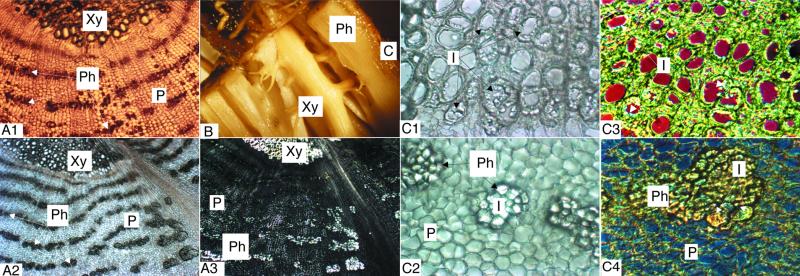
Figure 6.
Photographs showing fructan localization in T. officinale roots. A, Transverse cryosections of T. officinale root vascular tissue (magnification, ×22). Light microscopic view of an FAA- (A1) or 11.73 m ethanol fixed section (A2) and a section as in A2 but photographed under polarized light (A3). B, Anatomy of a fresh T. officinale root. Xylem and phloem can be easily separated from each other. C, Localization of inulin in xylem (C1 and C3) and phloem (C2 and C4) from the vascular tissue of a T. officinale root. Transverse cryosections (magnification, ×55). All tissues were fixed in 11.73 m ethanol for 2 d. Tissue slices 1 and 2 were photographed under a light microscope. Inulin crystals light up in the lignified metaxylem vessels (C1) and in clusters close to and surrounding the phloem tissue (C2). Figures C3 and C4 were obtained using a differential interference contrast microscope. Again, inulin crystals occupy the edge of the xylem vessels (C3) or appear as clusters surrounding the phloem (C4). This technique shows the crystals as striped structures marked by a color depending on the wavelength used. For all photographs: C, cortex; I, inulin; P, phloemparenchyma; Ph, phloem; Xy, xylem. All pictures were taken from a flowering T. officinale root.

