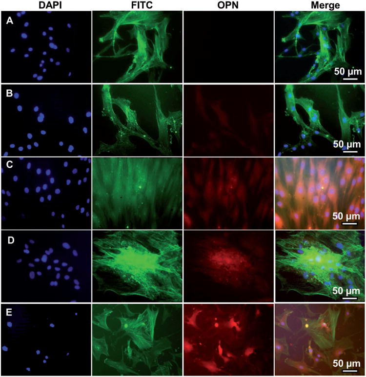
Figure 3.

Representative immunofluorescence images of OPN expression in human MSCs seeded on A) tissue culture plate (TCP) (A), flat (B, D), and nanoridged (C, E) SF films cultured in osteogenic induction-free medium (B, C) and osteogenic induction medium (D, E) on Day 14. The cells cultured on the nanoridged SF films (C) with osteogenic induction-free medium expressed more OPN than those cultured on TCP (A) and flat SF films (B) in the osteogenic induction-free medium. The cells cultured on both the flat (D) and nanoridged (E) films expressed OPN in the osteogenic induction medium. OPN was stained by rhodamine-labeled antibody (red) and cell nuclei were stained by 4′, 6-diamidino-2-phenylindole (DAPI) (blue) and F-actin was stained by FITC-labeled phalloidin (green).
