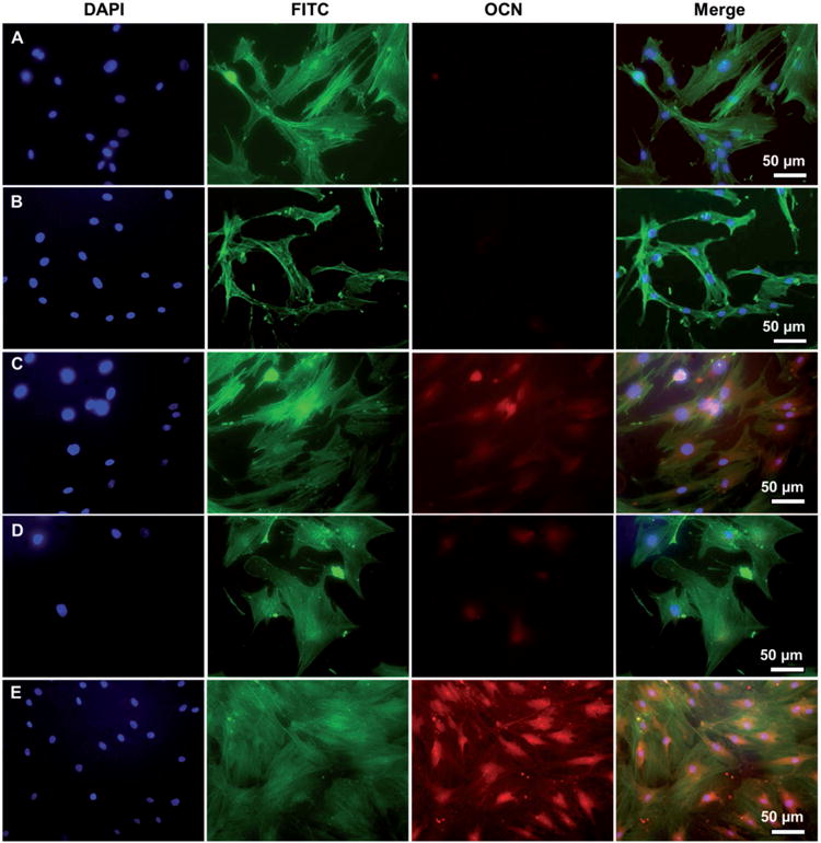
Figure 4.

Representative immunofluorescence images of OCN expression in human MSCs seeded on TCP (A), flat (B, D) and nanoridged (C, E) SF films cultured in osteogenic induction-free medium (B, C) and osteogenic induction medium (D, E) on Day 14. The cells cultured on the nanoridged SF films (C) with osteogenic induction-free medium expressed OCN, only slightly less than those cultured on the nanoridged SF films (E) with osteogenic induction medium. Moreover, cells cultured on TCP (A) and flat SF film (B) with osteogenic induction-free medium did not express OCN. Blue: DAPI nuclear staining; green: FITC actin staining; red: rhodamine-labeled antibody (red).
