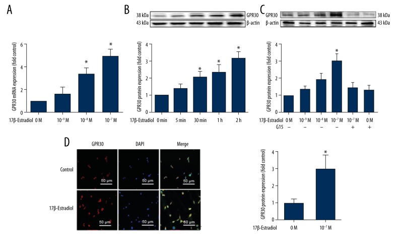
Figure 1.
Expression of the G-protein coupled estrogen receptor (GPER/GPR30) stimulated by 17β-estradiol in serum-starved ATDC5 chondrocytes. (A) Expression of GPER/GPR30 mRNA in serum-starved ATDC5 chondrocytes treated with increasing concentrations of 17β-estradiol (0 μM, 10−3 μM, 10−2 μM, and 10−1 μM), as assessed with quantitative real-time polymerase chain reaction (RT-PCR). (B) Expression of GPER/GPR30 protein in ATDC5 chondrocytes treated with 17β-estradiol (0 μM, 10 1 μM) for 0, 5, 30, 60, or 120 min, as assessed with Western blot. (C) Expressions of GPER/GPR30 protein in serum-starved ATDC5 chondrocytes treated with increasing concentrations of 17β-estradiol (0 μM, 10−3 μM, 10−2 μM, and 10−1 μM) with or without 15 μM of G15, as assessed by Western blot. (D) Confocal immunofluorescence (IF) staining of ATDC5 chondrocytes for GPER/GPR30 (red). The cell nuclei are stained with 4′, 6-diamidino-2-phenylindole (DAPI) (blue). The intensity of the GPER/GPR30 staining is increased in the serum-starved ATDC5 cells that were cultured in serum-free medium with 17β-estradiol (P <0.05). The figure data represent experiments performed in triplicate. The asterisks indicate significant differences compared with the control (* P<0.05).

