Figure 3.
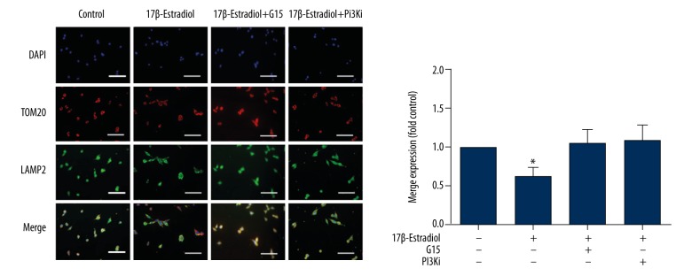
Confocal immunofluorescence (IF) staining for TOM20 and Lamp2 of ATDC5 cells. Confocal immunofluorescence staining for TOM20 (red) and Lamp2 (green). The cell nuclei are stained blue with 4,6-diamidino-2-phenylindole (DAPI). Serum-free ATDC5 cells were treated with 17β-estradiol (0 μM and 10−1 μM) with or without 15 μM of G15 and 20 μM of PI3K inhibitor. The staining intensity is lower in the serum-free 17β-estradiol-treated ATDC5 cell group compared with the other groups (P<0.05). White bar=50 μm.

