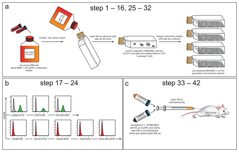
Figure 2. BM-MSC isolation, propagation and phenotypic characterization.
This figure and figure legend has been slightly modified from its original publication in: A humanized bone marrow ossicle xenotransplantation model enables improved engraftment of healthy and leukemic human hematopoietic cells. Nat Med 22, 812-821, doi:10.1038/nm.4103 (2016).25 [AU: Step numbers in figure and legend need updating – we can request the copy editors modify the step numbers in the figure if you so wish to avoid you needing to upload this figure again?]
(a) Experimental schematic describing isolation and propagation of human BM-derived MSCs (corresponding to STEP 1 – 9 and 18 – 25). Human total BM cells (from fresh BM aspirates or bag washouts) are admixed with expansion medium consisting of alpha modified minimum essential medium (alpha MEM) supplemented with 10% pooled human platelet lysate (pHPL)47,67 and seeded into cell culture vessels. After 24 to 48 hours non-adherent cells are removed by rinsing with pre-warmed PBS and the adherent cells are further expanded until the outgrowth of colonies (colony unit forming fibroblasts, CFU-F) is observed. These cells are considered passage 0 (p0). All p0 cells are detached from the plastic and sub-passaged into multiple flasks or cell factories to provide enough culture surface area to allow for sufficient expansion. Expanded cells (p1) are frozen or immediately used for the generation of humanized ossicle niches. (b) Purity of isolated MSCs is analyzed by flow cytometry using a consensus panel of positive and negative markers (corresponding to STEP 10 – 17).49 Histograms show representative cell surface staining for CD90, CD73 and CD105 (green) compared to isotype control (grey). BM-MSCs do not express hematopoietic markers CD45, CD14, CD34, CD19, or HLA-DR (red). X-axis shows mean fluorescence intensity (MFI) and y-axis shows counts. (c) Experimental schematic describing subcutaneous BM-MSC transplantation (corresponding to STEP 26 – 36). 2 × 10⁶ BM-MSCs are re-suspended in 60 µl of pHPL and admixed with 240 µl of extracellular matrix (Angiogenesis assay kit, Millipore) prior to subcutaneous injection (total volume: 300 µl per injection) into the flanks of 6 – 8 week old female NSG mice.

