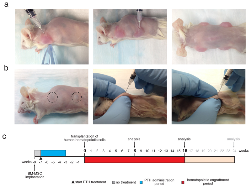
Figure 3. Humanized ossicle niche formation. [AU: Update step numbers.].
(a) Representative images (corresponding to STEP 33 - 35) showing subcutaneous injection of cell-matrix mixtures into the left front (left image) and left back (middle image) flank of a shaved NSG mouse. Up to 4 injections were done per mouse (right image). (b) Representative images (corresponding to STEP 38) showing humanized ossicle-niches formed 8 weeks after MSC transplantation visible through the skin of shaved mice (left image). Dashed circles highlight a purple hue indicative of proper ossicle niche formation and murine hematopoietic engraftment. Humanized ossicle-niches are readily accessible for direct intra-ossicle transplantation of human hematopoietic cells (corresponding to STEP 40 – 46) or ossicle marrow aspiration (corresponding to STEP 49) (middle and right image). (c) Experimental timeline. Starting at 3 – 7 days after BM-MSC transplantation (week -7) daily treatment of anabolic doses (40 µg/kg BW) of human 1-34 parathyroid hormone (PTH) is carried out for 28 consecutive days. Ossicle formation is evaluated visually (color change to purple indicates hematopoietic engraftment and BM niche formation, see dashed lines f, left image) weekly starting at week -2. Human hematopoietic cells (HSPCs, AML) are transplanted and engraftment is analyzed subsequently at indicated time-points up to 24 weeks post transplantation. All mouse experiments were conducted in accordance with a protocol approved by the Institutional Animal Care and Use Committee (Stanford Administrative Panel on Laboratory Animal Care no. 22264) and in adherence with the US National Institutes of Health’s Guide for the Care and Use of Laboratory Animals.

