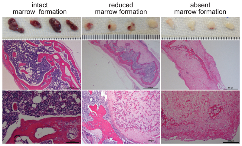
Figure 4. In vivo ossicle formation.
Examples of humanized organoids formed 8 – 10 weeks after BM-MSC transplantation into NSG mice as described in the protocol. Gross macroscopic images of all four generated ossicles harvested from one mouse (top) and overview and high power microscopic images of hematoxylin and eosin stains performed on the same ossicles. Scale bar indicate a millimeter scale for gross macroscopic image, 500µm for overview and 100µm (left image) or 200µm (middle and right image) for high power images. Most BM-MSCs successfully undergo endochondral ossification and form an intact marrow cavity populated by hematopoietic cells (left panel). Reduced or absent ossification can cause limited or absent marrow formation (middle and right panel). All mouse experiments were conducted in accordance with a protocol approved by the Institutional Animal Care and Use Committee (Stanford Administrative Panel on Laboratory Animal Care no. 22264) and in adherence with the US National Institutes of Health’s Guide for the Care and Use of Laboratory Animals.

