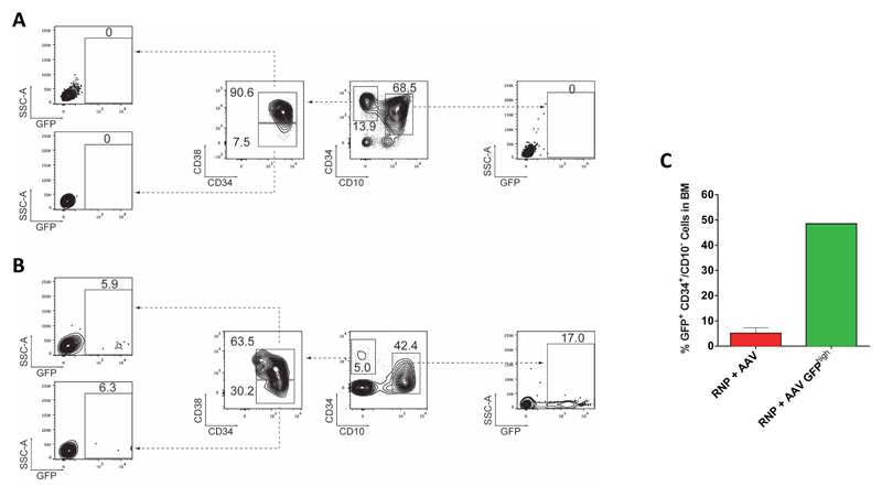
Extended Data Figure 7. Genome-edited human HSPCs in the bone marrow of NSG mice at Week 16 post transplantation.
Representative FACS plots from the analysis of NSG mice from the (A) Mock or (B) RNP+AAV experimental group at Week 16 post transplantation. Mice were sacrificed and bone marrow was harvested, PBMCs were isolated via Ficoll density gradient, after which human CD34+ cells were enriched by magnetic-activated cell sorting (MACS), and finally cells were stained with antiCD34, antiCD38, and antiCD10 antibodies to identify human GFP+ cells in the CD34+/CD10- and CD34+/CD10-/CD38- populations (note that CD10 was included as a negative discriminator for immature B cells) (C) Collective data from the analysis of GFP+ cells in the human CD34+/CD10- population from the RNP+AAV (N=11) and RNP+AAV GFPhigh (N=6) experimental groups. For the RNP+AAV GFPhigh group, cells from all six mice were pooled before analysis and thus, no error bar is available. Error bar on RNP group represents S.E.M.

