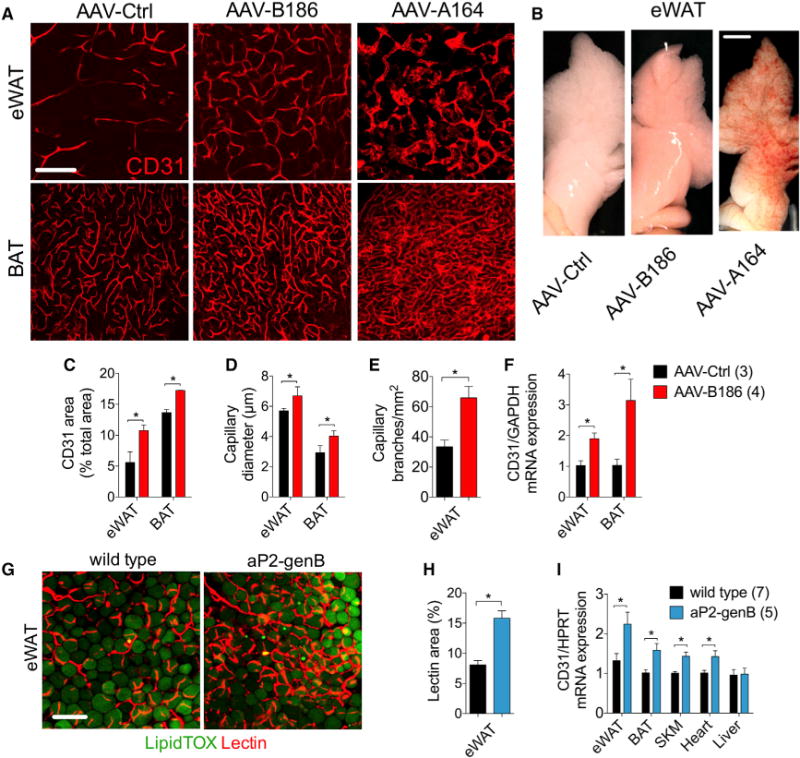
Figure 2. VEGFB Transduction Leads to Adipose Tissue Capillary Bed Expansion.

C57BL/6JOlaHsd males on a standard diet, transduced for 2 to 4 weeks with the indicated AAVs or transgenic for VEGFB were used for the experiments.
(A) Representative confocal projections of adipose tissue whole-mount staining for CD31.
(B) Macroscopic images of eWAT.
(C–E) CD31 area (C), capillary diameter (D), and capillary branch density (E) were quantified from images represented by the panels in (A).
(F) CD31 mRNA expression in adipose tissue.
(G and H) Representative confocal projections of adipose tissue whole-mount staining from mice injected intravenously with lectin (G) and lectin area quantification (H).
(I) CD31 mRNA expression in various tissues.
Scale bar, (A and G) 100 μm and (B) 3 μm. The number of mice is indicated in the figure. Data are represented as mean ± SEM. *p < 0.05, calculated with two-tailed unpaired t test.
