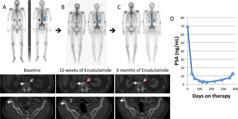
Figure 1.

(A) 99mTc-MDP skeletal scintigraphy and diagnostic CT performed at baseline, (B) after 10 weeks, and (C) after 6 months of enzalutamide in a 71-year-old man with known CRPC, who had previously been naïve to treatment with a second generation anti-androgen. Osseous disease at baseline became more sclerotic/conspicuous on the follow-up bone scan and CT after 10 weeks of therapy (white arrows) correlating with decreasing PSA (D). The 6 month follow-up bone scan and CT confirms a flare phenomenon, similar to that described in the literature1–4. There was also lymph node disease on the baseline diagnostic CT (pink arrows) that improved on the diagnostic CT scans obtained after 10 weeks and 6 months of therapy. PSA nadir was seen at approximately 6 months post-initiation of enzalutamide, and therapy was discontinued at 1 year due to progression.
