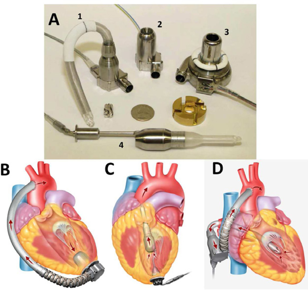
Figure 4.
The HeartWare HVAD (A #3) and various configurations of the HeartWare MVAD in development (A #1, 2, 4) are shown. The first transapical MVAD configuration is implanted through the left ventricular apex (A #2, B) with the outflow graft sewn to the descending aorta. The second, transapical MVAD configuration is implanted with a subxiphoid approach (A #4, C). The transmitral MVAD configuration is implanted through a small, right-sided thoracotomy (A #1, D).

