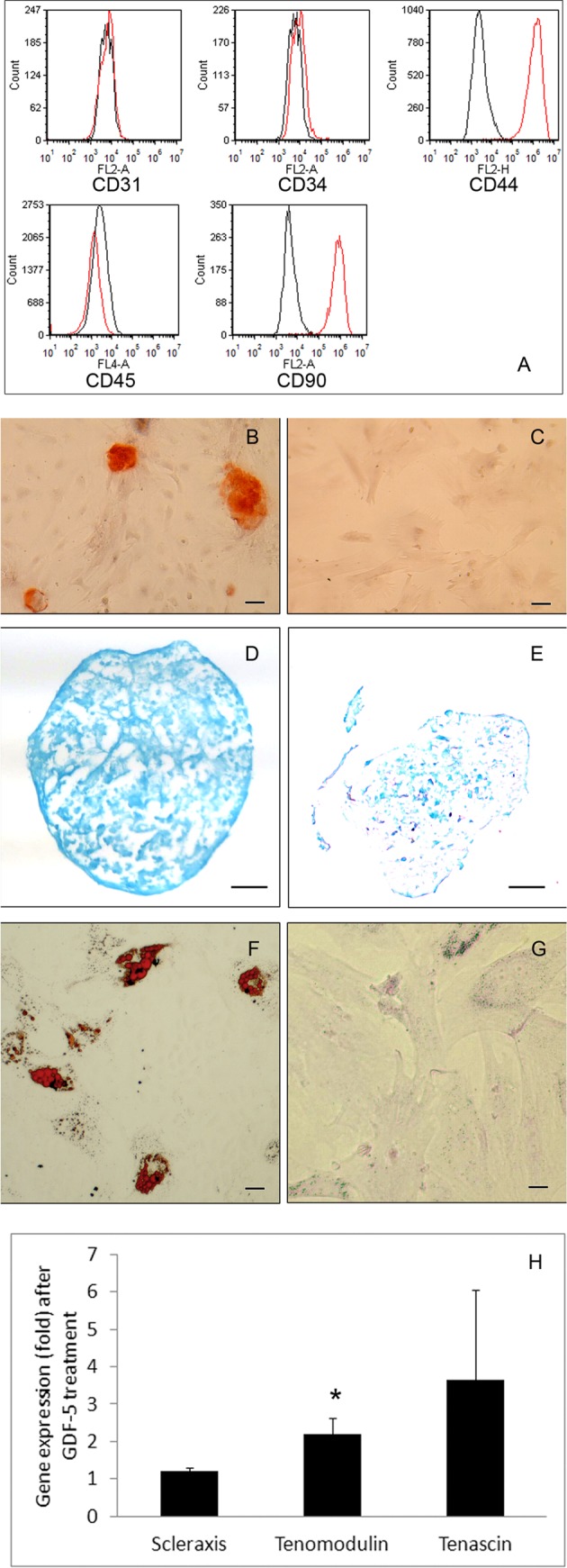
Fig. 1.

Characterization of adipose tissue–derived mesenchymal stem cells (MSCs). A panel of cell surface markers examined by Fig. 1. (continued). flow cytometry (A). The adipose tissue–derived MSCs do not express CD31, CD34, and CD45 but express CD44 and CD90. In osteogenic cultures, there is mineralization in the extracellular matrix (red; B) but not in the control cultures (C; Alizarin Red staining; bar = 50 µm). Chondrogenic pellet cultures show abundant proteoglycans (blue; D), but the control pellet (E) has little proteoglycans in the matrix (Alcian Blue staining; bar = 100 µm). In adipogenic cultures, fat droplets are developed intracellularly (red; F) but not in the control cultures (G; Oil Red O staining; bar = 20 µm). The expression of scleraxis and tenascin by MSCs treated with or without growth differentiation factor-5 (GDF-5) is not different statistically (H). The expression of tenomodulin by MSCs treated with GDF-5, however, is significantly increased over the control (P < 0.05).
