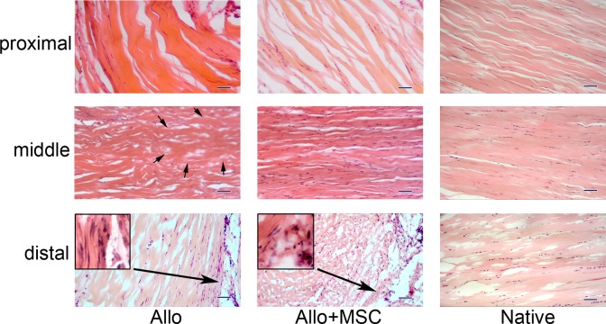
Fig. 4.
Histology of the transplanted Achilles allograft, with or without mesenchymal stem cells (MSCs). For both Allo and Allo + MSC groups, there are fibroblasts invaded in the proximal portion of the allograft, and some of the cells appear elongated and tenocyte-like. In the middle segment of the allograft, while acellular area (outlined with arrows) exists in the Allo group, the allograft is fully repopulated in the Allo + MSC group. There are abundant fibroblasts that infiltrated into the distal portion of the allograft in both groups, particularly at the peripheral region (inserts in higher magnifications). The corresponding proximal, middle, and distal portions of native Achilles tendon are posted for control (hematoxylin and eosin staining; bar = 100 µm).

