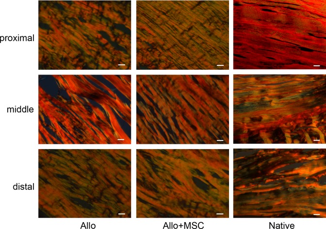
Fig. 5.
Collagen composition in Achilles allograft by polarizing microscopy. The allograft in both Allo and Allo + mesenchymal stem cell (MSC) groups and native tendons are overwhelmingly stained for thick (type I) collagen fibers (red) in all 3 segments. Compared with the native Achilles tendon, the proximal and distal portions of the allograft in both Allo and Allo + MSC groups have increased deposition of fine (type III) collagen fibers (green). In the middle portion of the allograft, while the deposition of fine collagen fibers mostly appear around the large collagen bundles in the Allo group, it takes place inside of the collagen bundles in the Allo + MSC group (picrosirius Red staining; bar = 50 µm).

