Figure 3.
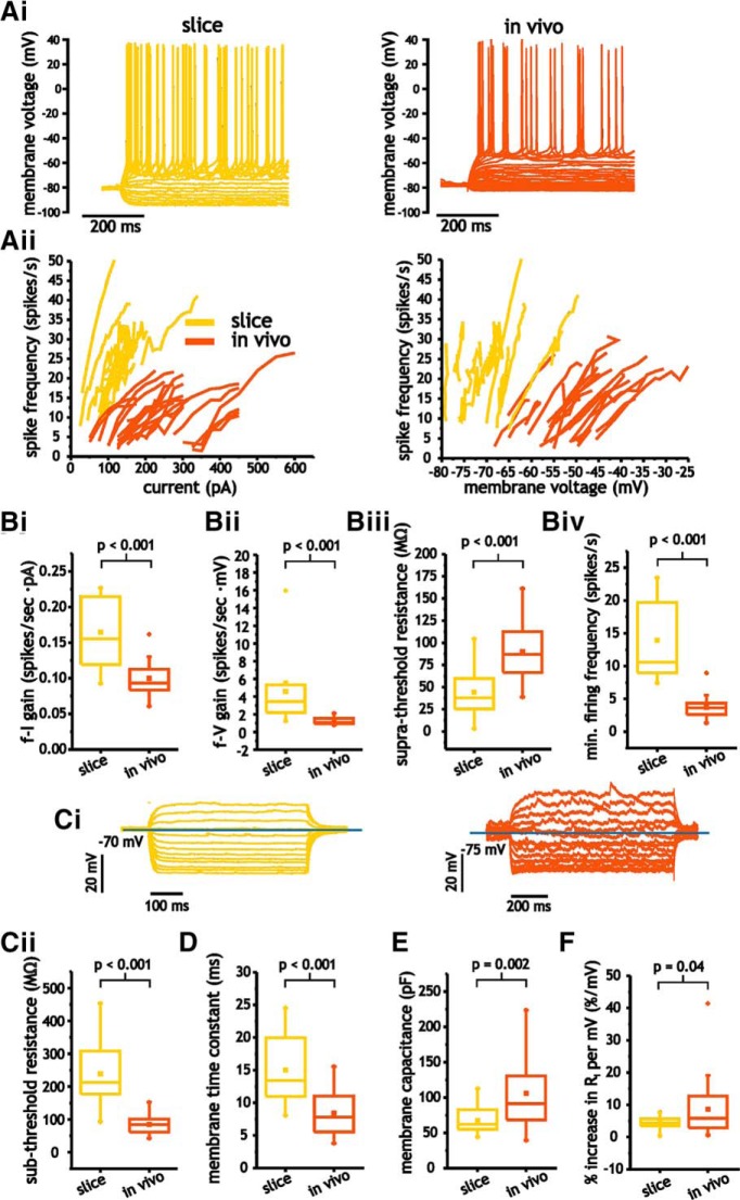
Pyramidal cells express significant differences in electrophysiological properties between in vivo and slice conditions. A, Representative voltage traces from pyramidal cells in response to step depolarizations (i) and f-I and f-V curves (ii) for both in vivo and slice neurons. B, Comparison of average f-I (i) and f-V (ii) gain, as well as suprathreshold membrane resistance (iii) and minimum spike firing frequency (iv) in pyramidal cells in vivo and in slices. Ci, Representative subthreshold voltage traces from pyramidal cells in response to step depolarizations in vivo and slice neurons. Cii–E, Comparison of average membrane input resistance (Cii), time constant (D), and membrane capacitance (E) in pyramidal cells under in vivo and slice conditions. F, Comparison of average percentage increase in subthreshold membrane resistance in vivo and slice conditions.

