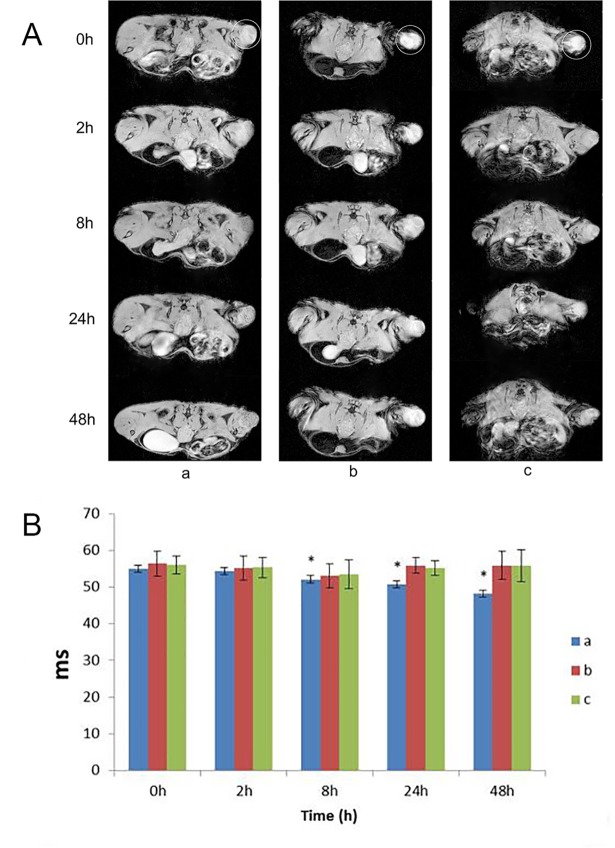
Fig 3.
The MRI T2*WI of the human transplanted glioma in mice labeled by Fe3O4@Au-C225 composite targeted MNPs(a), Fe3O4@Au composite MNPs(b), both Fe3O4@Au-C225 composite targeted MNPs and C225(c) (circle for the glioma)(A); The T2*WI relaxation time of the human glioma transplanted in nude mice labeled by Fe3O4@Au-C225 composite targeted MNPs(a), Fe3O4@Au composite MNPs(b), both Fe3O4@Au-C225 composite targeted MNPs and C225(c) at different time points (*compared with the 0h time point, P<0.05)(B).

