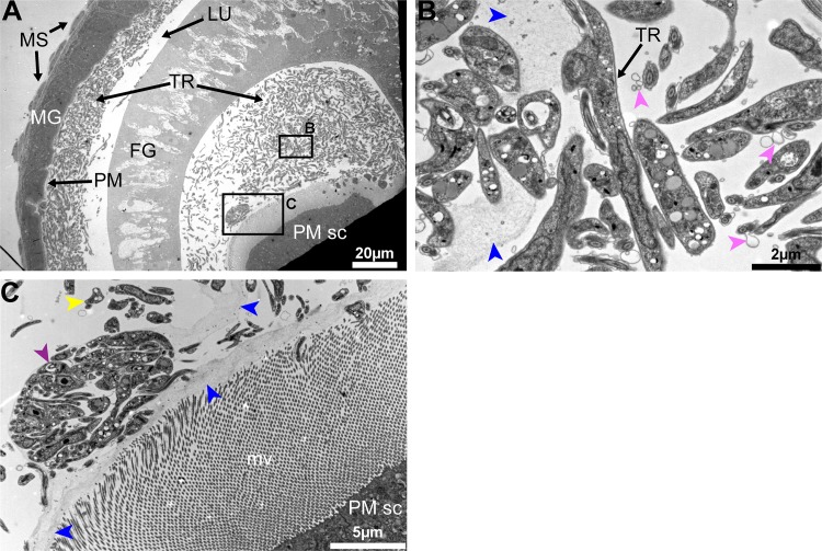
Fig 4. Trypanosome-PM interactions in cardia from inf+/+ tsetse.
(A-C) Ultrastructure of PM secreting cells in the cardia from inf+/+ flies. (A) Trypanosomes are observed in mass in the lumen. (B-C) Magnified micrographs of the black boxes shown in (A). (B) Trypanosomes are observed embedded in the secreted matrix (blue arrowheads). (C) In this niche parasites are observed in cyst-like bodies (purple arrowhead), and can also be observed out of the PM secretions (yellow arrowheads). Parasite secreted extracellular vesicles are observed (pink arrowheads). Micrographs in this image represent one of six biological replicates analyzed. MG: midgut; FG: foregut; TR: trypanosomes; MS: muscles; PM: peritrophic matrix; PM sc: PM secreting cells; mv: microvilli.

