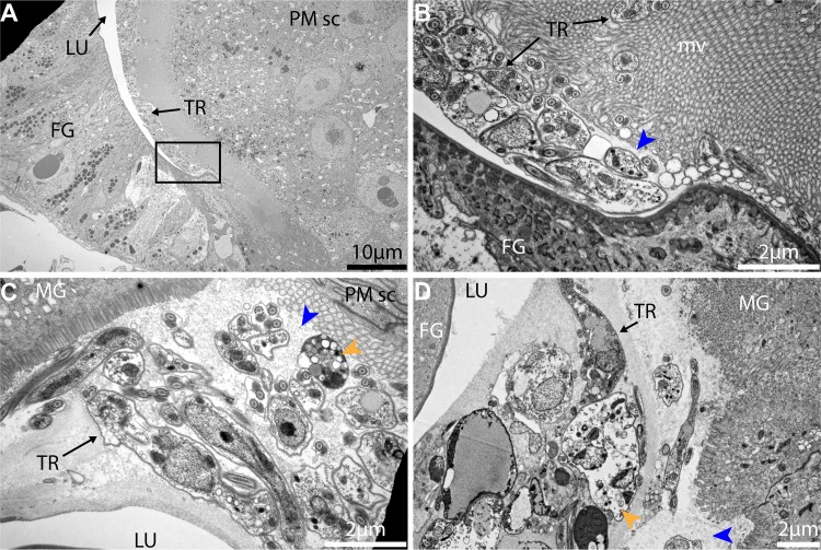
Fig 5. Trypanosome-PM interactions in cardia from inf+/- tsetse.
(A-D) Ultrastructure of cardia inf+/- near the PM secreting cells. (B) is a magnification of the black frame in (A) showing the PM (blue arrowhead). (C) and (D) two independent cardia organs showing the same region near PM secreting cells. Trypanosomes are observed packed within the ES near the location of PM secretion. At this point, several trypanosomes observed present vacuolation and nuclear condensation (orange arrowheads) indicative of cell death. Micrographs in this image represent three of five biological replicates analyzed. MG: midgut; FG: foregut; TR: trypanosomes; PM: peritrophic matrix; PM sc: PM secreting cells; mv: microvilli.

