
Figure 1. Uterine artery Doppler velocimetry findings in the second trimester of pregnancy.
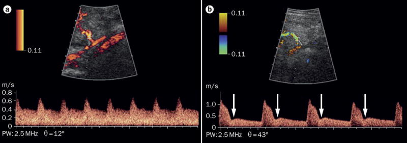
A. Normal findings. B. Abnormal findings, indicated by either the presence of bilateral uterine artery early diastolic notches (arrows) or a mean pulsatility index (calculated as [peak systolic velocity – end diastolic velocity]/time averaged velocity, averaged across both uterine arteries), above the 95th percentile for gestational age.