
Figure 1.
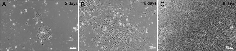
Nesting of mouse pulmonary microvascular ECs. Phase contrast micrographs of mouse pulmonary microvascular endothelial cells culture on (A) day-2 showing Dynabeads® in culture and an inset with a “nest” of endothelial cells and; (B) day-6; (C) Shows a phase-contrast confluent monolayer on day-8. Scale bars are 200 μm.