
Figure 3.
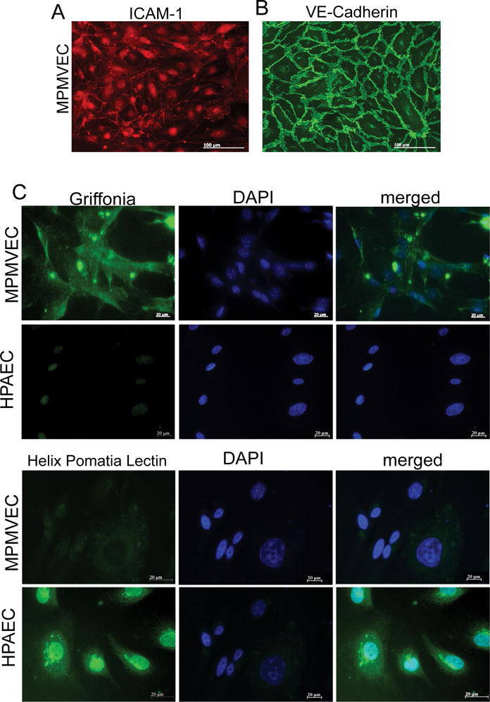
A Characteristics of mouse pulmonary microvascular endothelial cells (MPMVECs). MPMVECs were cultured at 37 °C in complete endothelial growth basal medium-2 on fibronectin until confluent (A) Expression of Intercellular adhesion molecule-1 (ICAM-1) in representative micrograph of cultured endothelial cells; (B) Expression of VE-cadherin is localized at the cell-cell junctions shown in representative micrograph with expression of vascular endothelial cadherin; (C) Endothelial cells isolated from murine lungs or HPAEC stained either with Griffonia or Helix Pomatia lectin. Scale bars are 20 μm.