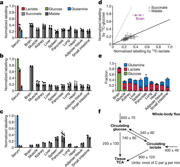
Figure 2. In fasting mice, glucose labels TCA intermediates through circulating lactate in all tissues except the brain.

a–c, Normalized labelling of serum glucose, lactate, and glutamine, and of tissue TCA intermediates by infused 13C-lactate (a, n = 4), 13C-glucose (b, n = 5), and 13C-glutamine (c, n = 3). Data are mean ± s.d. d, Scatter plot of normalized labelling of TCA intermediates by infused 13C-glucose versus infused 13C-lactate (13C-glucose and 13C-lactate experiments performed separately). The solid line represents the expected labelling by 13C-glucose assuming that glucose feeds the TCA cycle solely through circulating lactate. The dashed line indicates the expected labelling by 13C-lactate assuming that lactate feeds the TCA cycle solely through circulating glucose. Data are from a and b, each data point is one TCA intermediate in one tissue, mean ± s.d., n = 4 for 13C-lactate infusion and n = 5 for 13C-glucose infusion. e, Direct circulating nutrient contributions to tissue TCA cycle (see Supplementary Note 3), data are mean ± s.e.m. f, Steady-state whole-body flux model of interconversion between circulating glucose and lactate and their feeding of TCA (see Supplementary Note 4), data are mean ± s.e.m.
