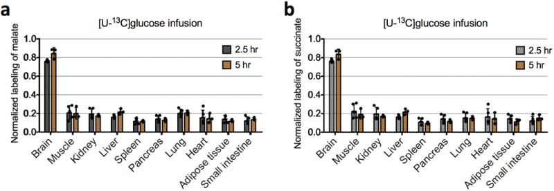
Extended Data Figure 4. Isotopic labelling of tissue TCA intermediates reaches steady state after 2.5-h infusion of 13C-glucose.

a, Comparison of normalized labelling of tissue malate after 2.5 h (n = 5 mice; mean ± s.d.) and after 5 h of [U-13C]glucose infusion (n = 3 mice; mean ± s.d.). P values were determined by an unpaired Student’s t-test, corrected for multiple comparisons using the Holm–Sidak method. Normalized labelling is the fraction of 13C atoms in a metabolite divided by the fraction of 13C atoms in serum glucose. None of the differences are significant (P > 0.14 for the brain and liver, and P > 0.98 for other tissues). b, Comparison of normalized labelling of tissue succinate after 2.5 h (n = 5 mice; mean ± s.d.) and after 5 h of [U-13C]glucose infusion (n = 3 mice; mean ± s.d.). None of the differences are significant (P > 0.18 for the brain and liver, and P > 0.85 for other tissues).
