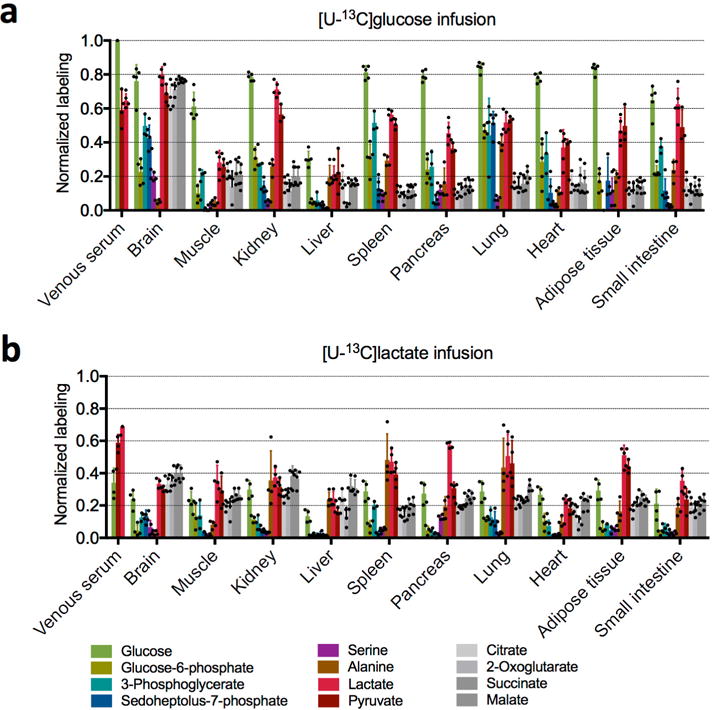
Extended Data Figure 5. Isotope labelling of central carbon metabolites by 13C-glucose and 13C-lactate.

a, Normalized labelling by 13C-glucose in fasting mice (n = 3 for pyruvate, 2-oxoglutarate, and 3-phosphoglycerate, n = 4 for alanine, and n = 5 for all other metabolites; mean ± s.d.). b, Normalized labelling by 13C-lactate in fasting mice (n = 3 for 3-phosphoglycerate and n = 4 for all other metabolites; mean ± s.d.). n indicates the number of mice. For the lactate tracer studies, venous serum and tissue labelling are normalized to the arterial serum lactate labelling. Note that citrate, malate and succinate in tissues turn over sufficiently slowly that labelling is robust to the small (<90 s) delay between euthanizing the mouse and tissue harvesting. This delay may, however, result in erroneous measurements for tissue lactate, pyruvate and glycolytic intermediates. Analyses in the main text are limited to the better validated measurements of serum metabolites and tissue TCA intermediates. With this caveat in mind, it is nevertheless intriguing that lactate labelling varies markedly across tissues. After labelled glucose infusion, lactate labelling is highest in the brain, consistent with its use of glucose as a major substrate. In the kidneys lactate is strongly labelled after glucose infusion, even though TCA intermediates are more labelled after lactate infusion. A potential explanation involves tissue heterogeneity; for example, the presence both of glycolytic cells that make lactate from circulating glucose and of oxidative cells that make TCA intermediates from circulating lactate. In other tissues, such as liver, tissue lactate labelling is far below circulating lactate and very similar to TCA labelling; this may reflect mixing of carbon between lactate and TCA intermediates via gluconeogenesis or pyruvate cycling. Another factor diluting tissue lactate labelling is that, as blood passes through tissue, owing to the rapid exchange between tissue and circulating lactate, the circulating lactate loses its labelling, as is evident from the lactate arteriovenous labelling difference.
