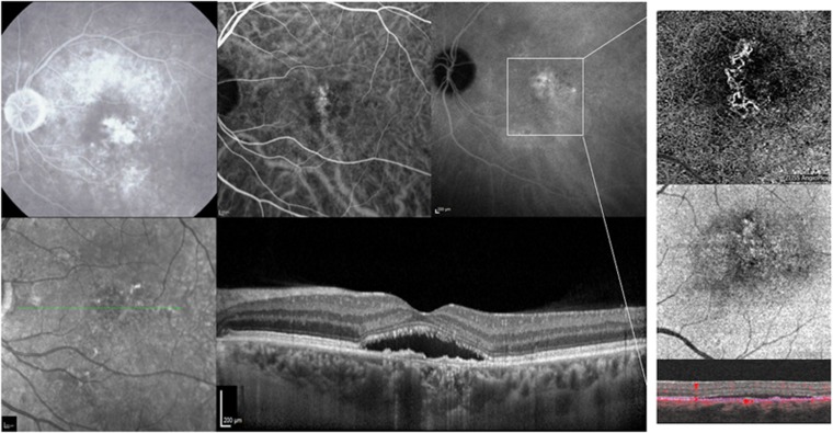
Figure 3.
Fluorescein angiography (FA), indocyanine green angiography (ICGA), infrared reflectance (IR), optical coherence tomography (OCT), and OCT-angiography (OCT-A) of a patient affected by central serous chorioretinopathy (CSC) from 4 months. (Top row, first panel) FA at 5 min postinjection revealing a late-phase ill-defined hyperfluorescent lesion, without leakage or pooling of dye. (Top row, second and third panel) Early and late ICGA images showing a hyperfluorescent area corresponding to the choroidal neovascularization (CNV) along with punctate hyperfluorescent spots. (Bottom row, first and second panel) IR and structural OCT B-scan passing through the fovea showing the presence of a macular serous retinal detachment and the presence of a flat irregular elevation of the RPE with moderately reflective material in the sub-RPE space, compatible with a type 1 CNV. (Top, middle and bottom row, fourth panel) Choriocapillaris segmentation on 3 × 3 OCT-A, corresponding en-face OCT and B-scan with flow showing the presence of CNV.

