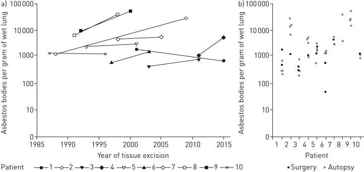
FIGURE 3.
Results of fibre analyses at surgery (first tissue excision) and autopsy (second tissue excision) in relation to time from 10 patients. From these 10 patients the asbestos fibre burden of the lung has been determined from tissue both times. Two patients with fibre analysis from a bronchoalveolar lavage are not considered here. Illustrated are the results of the highest asbestos body count in relation to time (a) and all asbestos body counts from one patient's lungs separately (b). The corresponding time interval is already illustrated in figure 2 and listed in table 1. It is easily seen in (b) that the counts from surgery are within the range of the counts from autopsy. For two patients the lower count from surgery tissue could be explained histologically (supplementary material). Patient 2 had tuberculosis in the first tissue and patient 6 had fibrosis with a multi-etiological clinical picture with an asbestos-dependent and an asbestos-independent component (see supplementary material for details).

