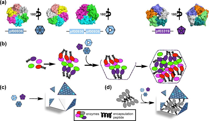
Figure 1.
Shell proteins (a) and assembly of BMCs (b–d). (a) Representatives of a BMC-H protein (PDB 5DJB) (left), a BMC-T protein (PDB 5DIH) (middle), and a BMC-P protein (PDB 2QW7) (right). Pf indicates Pfam identification. Individual polypeptide chains are colored differently. (b) Cartoon representation of BMC assembly from the inside out, where the primary role of the EP is in shell recruitment [25, 35••] (c) of empty BMC shell assembly, and (d) of targeting enzymes to the lumen of BMC shells using EPs [36••].

