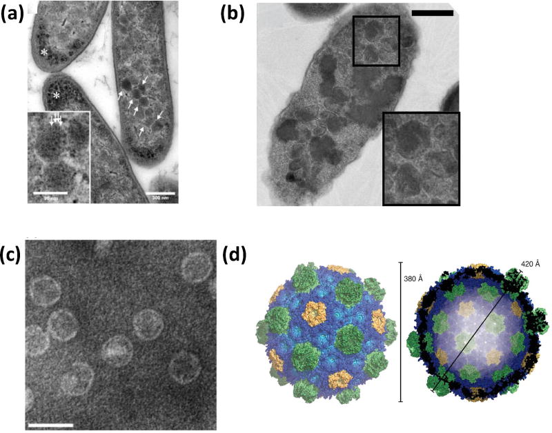
Figure 2.
Physical characterization of engineered BMCs. (a) Thin section electron micrograph of E. coli expressing the complete PDU BMC operon (arrows) (scale bar 300 nm). Asterisks mark unknown granular dense matter. Inset is an enlarged view of the polyhedral bodies (scale bar 96 nm) that show regular substructures (arrows) [37]. (b) Thin sections electron micrograph of E. coli expressing the carboxysome genes of Halothiobacillus neapolitanus viewed by TEM (scale bar 500 nm) showing polyhedral bodies (magnified in inset) [39]. (c) Negatively stained electron micrograph of purified HO BMC shells (scale bar 50 nm). Reproduced with permission from [40]. Copyright Elsevier 2014. (d) Surface representation of the HO shell crystal structure. Shell proteins are colored blue (BMC-H), green (BMC-T), and yellow (BMC-P). Reproduced with permission from [41••]. Copyright The Association for the Advancement of Science 2017.

