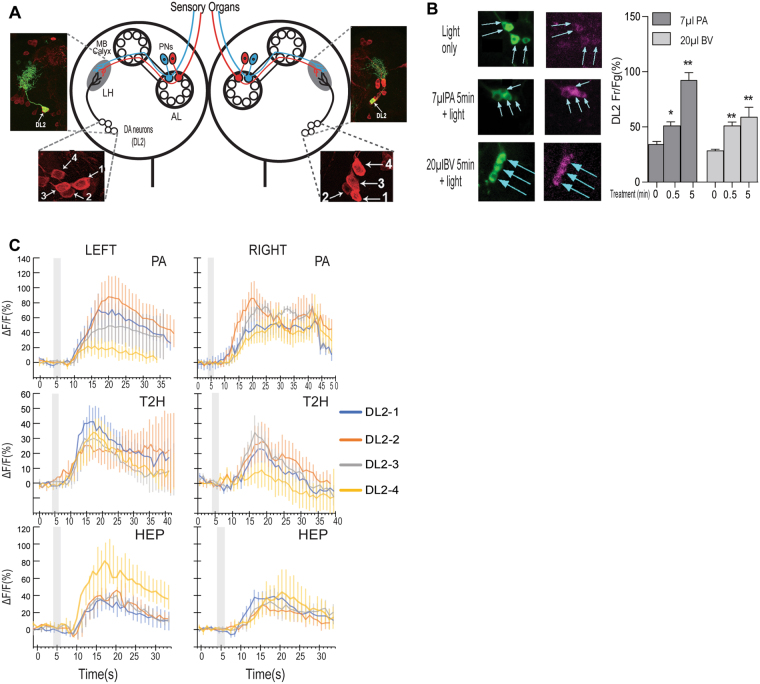
Figure 2.
Convergence of food odor inputs onto the assembly of DA neurons. (A) Schematic drawing of the larval olfactory circuit shows two clusters of four DA neurons (DL2-1 to 4) in the left and right brain hemisphere7. The DL2 neurons project dendrites to the lateral horn (LH) in each brain lobe, where they form synaptic connections with the projection neurons. Upper insets show that the axons and dendrites of individual DL2 neurons, labeled with mCD8::GFP, are exclusively located in the LH region. AL: antenna lobe; MB: mushroom bodies; PNs: projection neurons. DL: dorsolateral. (B) CaMPARI-based fluorescence imaging of freely behaving larvae revealed the dynamic responses of four DL2 neurons to an effective dose of PA or balsamic vinegar (BV) during 5-minute stimulation. For each time point, at least 29 DA neurons were imaged. Data are presented as ± SEM. *P < 0.05; **P < 0.001 (C) GcaMP6 imaging revealed that in fed larvae, DL2 neurons from the left and right brain hemisphere showed differential excitatory responses to each of the three monomolecular odorants. The shaded box indicates the duration of odor stimulation. In each imaging assay, 5–6 neurons were examined for each cell type.

