Figure 6.

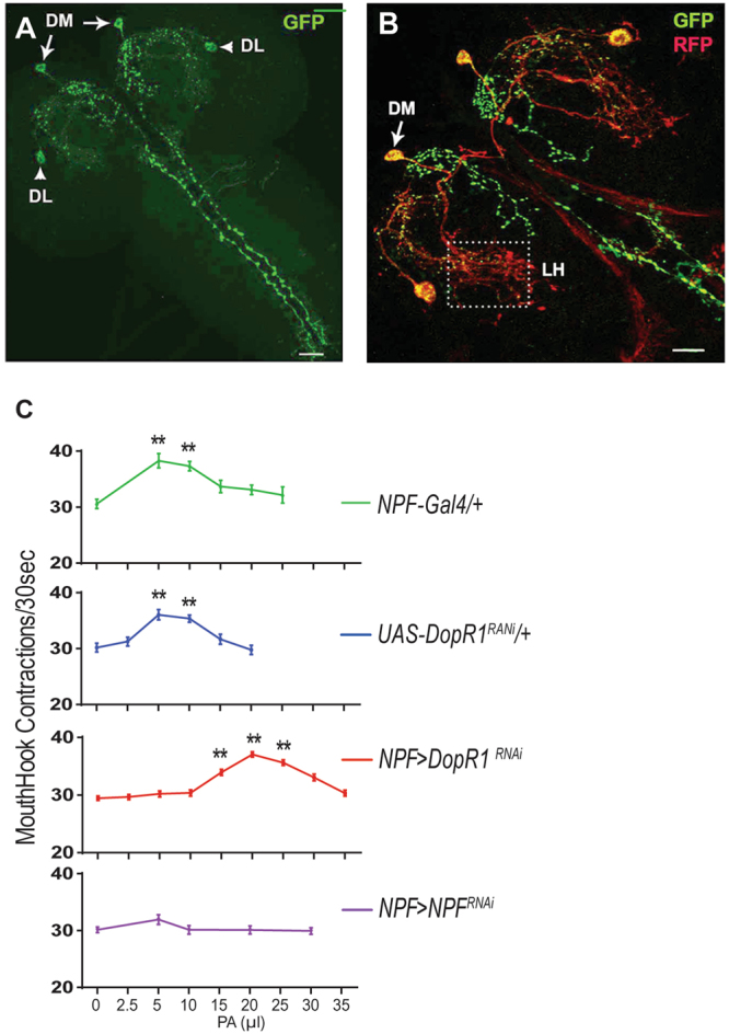
Mapping of the gating activity of Dop1R1 to NPF neurons. (A) An imaging of two pairs of neurons in the larval brain that are selectively labeled by npf-GAL4. DM: the dorsomedial pair; DL: the dorsolateral pair. (B) Expression of sytGFP in the axons (green) and denmark in the dendrite (red) of the npf-GAL4 neurons. There is an extensive presence of NPF neuronal dendrites in the lateral horn (LH). (C) The dose-response curves of Dop1R1-deficient, npf-deficient and control larvae (also see Figure S3 for other controls). n > 15; **P < 0.001.
