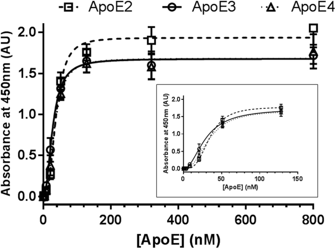
Figure 3.

Saturation binding of apoE isoforms to the ELISA polystyrene surface. Variable concentrations (0–800 nM) of the three different recombinant apoE isoforms were added to ELISA plate wells, and incubated for 2 h at RT. After blocking, bound apoE was detected with a polyclonal pan-apoE antibody, followed by horseradish peroxidase-labelled anti-rabbit IgG. Error bars represent the standard deviation of duplicated measures. The graph shows a representative experiment of four independent tests performed by duplicate. The inset shows the same data represented from apoE concentrations in the 0–128 nM range.
