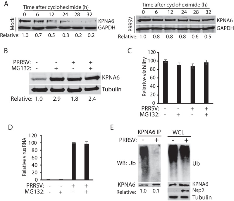
FIG 2.

PRRSV extends KPNA6 half-life and reduces KPNA6 polyubiquitination. (A) PRRSV infection extends KPNA6 half-life. MARC-145 cells were infected with VR-2385 at an MOI of 1. The cells were treated with cycloheximide at 24 hpi and harvested at the indicated time (h) for WB. Mock-infected cells at the corresponding time points were included as controls. The relative levels of KPNA6 are given below the images. (B) MG132 treatment blocks KPNA6 turnover in MARC-145 cells. The cells were infected with VR-2385 at an MOI of 1. At 18 hpi, the cells were treated with MG132 and, 6 h later, harvested for WB with antibodies against KPNA6 and tubulin. The MG132-negative wells were treated with the solvent, DMSO. Mock-infected cells were included as controls. The relative levels of KPNA6 are shown below the images after normalization with tubulin. (C) Cell viability assay of MARC-145 cells treated with MG132 or DMSO and/or infected with PRRSV. (D) PRRSV viral RNA level in cells treated with MG132 or DMSO. (E) PRRSV infection reduces KPNA6 polyubiquitination. MARC-145 cells were infected with PRRSV VR-2385 at an MOI of 1 and harvested for IP with KPNA6 antibody at 24 hpi, followed by WB with the ubiquitin (Ub) antibody. WB of the whole-cell lysate (WCL) with the antibodies against ubiquitin, KPNA6, PRRSV nsp2, and tubulin was conducted. The relative levels of Ub after normalization with KPNA6 are shown below the images.
