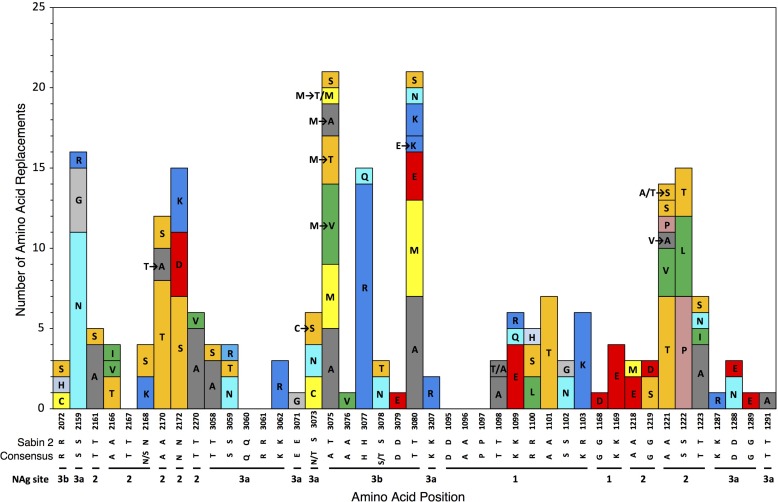
FIG 2.
Numbers of independent amino acid replacements in NAg sites of all 403 Nigerian VDPV2 variants isolated from 2005 to 2011. The positions are ordered starting from the N terminus of the capsid, aligned with the corresponding residues of Sabin 2 and the consensus among 12 isolates representing distinct WPV2 genotypes. Amino acids are colored by physicochemical properties according to the Amino/Shapely scheme (A [nonpolar, small aliphatic], dark gray; C and M [nonpolar, sulfur-containing], yellow; D and E [charged, acidic], bright red; F and Y [polar and nonpolar aromatic], mid-blue; G [nonpolar, no side chain], light gray; H [charged, imidazole], pale blue; I, L, and V [nonpolar, aliphatic], green; K and R [charged, basic], blue; N and Q [polar, amide], cyan; P [nonpolar, cyclic imino], flesh; S and T [polar, hydroxyl], orange; W [nonpolar, aromatic, indole], pink) (http://acces.ens-lyon.fr/biotic/rastop/help/colour.htm#shapelycolours).

