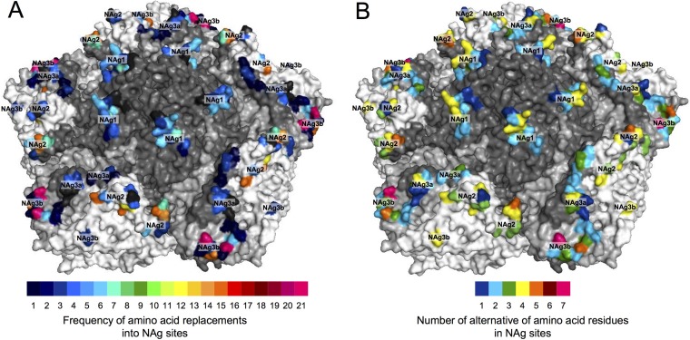
FIG 7.
Heat maps illustrating the frequency of NAg amino acid replacements (A) and the number of alternative amino acid residues (B) in the NAg sites of all 403 cVDPV2 isolates mapped to the X-ray crystallographic structure of the poliovirus type 2 capsid pentamer. Capsid proteins (see Fig. S7 in the supplemental material) are shaded as follows: VP1, dark gray; VP2, white; VP3, light gray.

