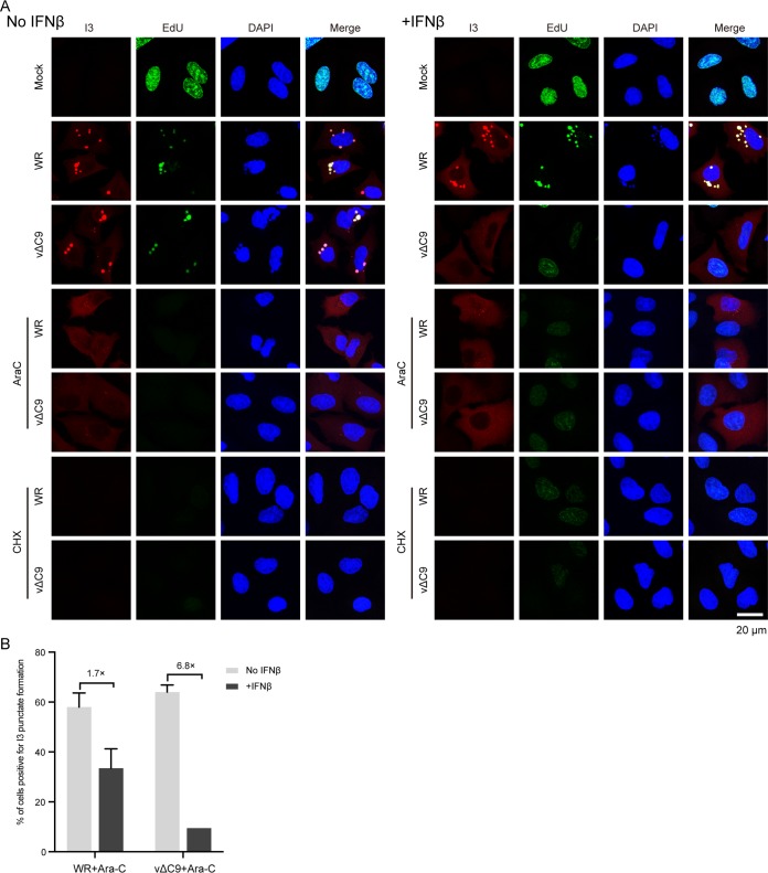
FIG 6.
Effect of IFN-β on release of the vΔC9 genome from viral cores. (A) Confocal microscopy. A549 cells were pretreated with 2,000 IU/ml of IFN-β for 24 h and then mock infected or infected with 3 PFU/cell of purified WR or vΔC9 in the presence or absence of AraC or CHX. After 3 h, the cells were incubated with 10 μM EdU for 1 h, fixed, and permeabilized. EdU was reacted with Alexa Fluor 647 azide by click chemistry, and the cells were stained with antibody to the I3 protein followed by Alexa Fluor 568 secondary antibody and DAPI and examined by confocal microscopy. Bar, 20 μm. (B) Automatic tiling was used to select fields of cells, and the percentages of AraC-treated cells positive for I3 punctate spot formation relative to the total number of cells expressing I3 were determined manually. Approximately 300 I3-positive cells were counted. The error bars represent standard deviations.

