Fig. 1.
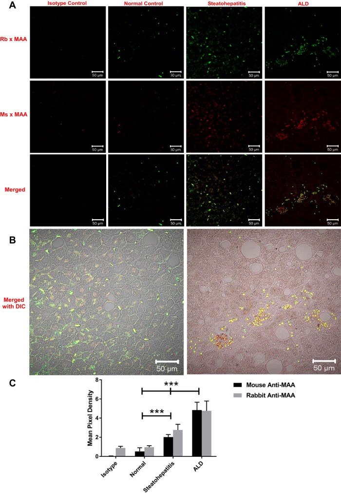
Immunohistochemical staining for malondialdehyde-acetaldehyde (MAA) adducts in human liver tissue. Paraffin-embedded liver sections from patients with normal liver, steatohepatitis, and alcoholic liver disease (ALD) were subjected to immunohistochemical staining using a rabbit (Rb) and a mouse (Ms) anti-MAA antibody. A: staining with rabbit anti-MAA antibody (green, top) and mouse anti-MAA antibody (red, middle) and merged images (yellow, bottom). B: merged differential interference contrast (DIC) images of steatohepatitis and ALD samples stained with both anti-MAA antibodies. C: quantification of mean pixel density. Sections were mounted in Fluoromount-G and viewed using a confocal laser-scanning microscope (Zeiss 710 Meta). Images were analyzed using Zen 2.1 Black (Zeiss) and ImageJ software. Values are means ± SE (n = 5). ***P < 0.001 vs. normal and steatohepatitis.

