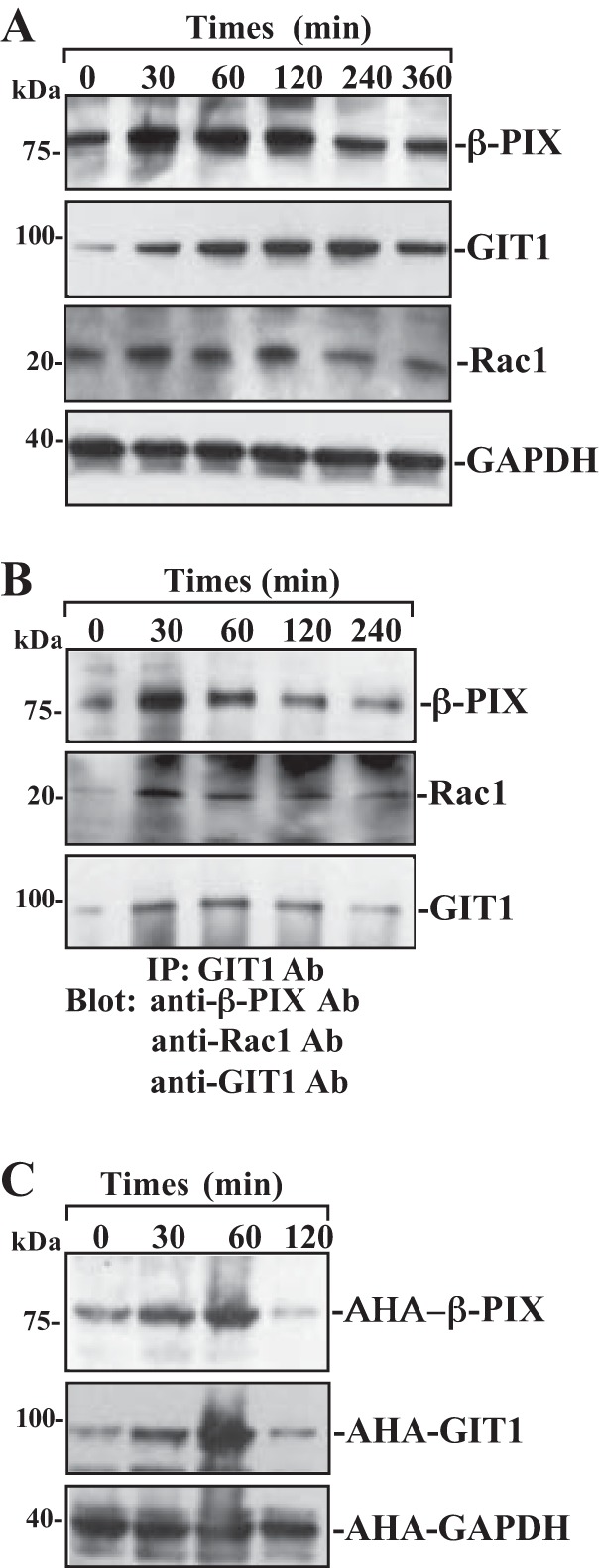
Fig. 2.

Changes in the levels of β-PIX, GIT1, Rac1, and β-PIX/GIT1/Rac1 association after wounding. After IEC-Cdx2L1 cells were grown to confluence, epithelial restitution was induced by removing part of the monolayer, as described in materials and methods. A: levels of β-PIX, GIT1, and Rac1 proteins in total cell lysates isolated at various times after wounding and detected by Western blot analysis. GAPDH immunoblotting was performed as an internal control for equal loading. B: levels of β-PIX, GIT1, and Rac1 proteins in IP materials by the anti-GIT1 Ab from the samples described in A. C: newly synthesized β-PIX and GIT1 proteins as measured by Click-IT protein analysis using l-azidohomoalanine (AHA) in samples described in A. Three experiments were performed that showed similar results.
