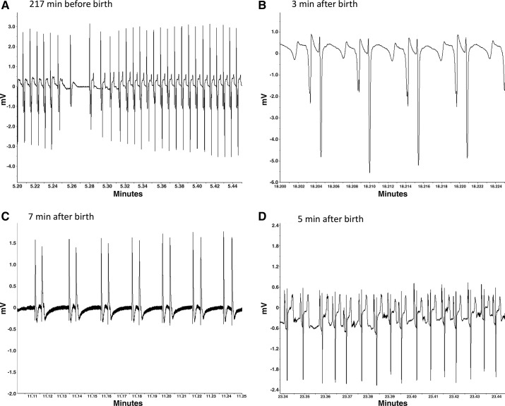
Fig. 5.
Examples of ECG abnormalities identified in four cortisol-treated fetuses or lambs at the time of birth. A: atrioventricular (AV) block (P wave with no associated QRS complex) and elevated ST segment in a fetus during active labor. B: notched (or split) P wave in a newborn lamb. C: atrial fibrillation in a newborn lamb who later succumbed. D: abnormal ST segment and notched P waves in a newborn lamb. Note that the shape of the ECG varies with the positioning of the lead on the chest wall relative to the lead secured within the jugular. Thus the shape can vary between fetuses and also within fetus, depending on the orientation of the fetus.

