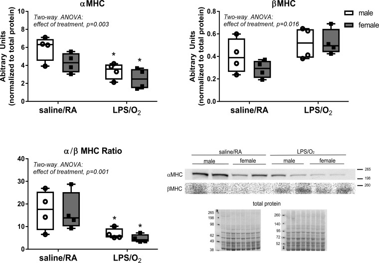
Fig. 2.
Myosin heave chain (MHC) isoforms were determined by Western blot. Left ventricular homogenates were separated by SDS-PAGE, transferred to PVDF membranes, and probed with anti-α-MHC or anti-β-MHC antibodies. Membranes were quantified by densitometry. Data were analyzed by two-way ANOVA with Tukey’s post hoc analyses. Two-way ANOVA identified an effect of treatment, α-MHC, P = 0.003, and β-MHC, P = 0.016, and individual differences were observed between the saline/room air (RA) and lipopolysaccharide (LPS)/O2 measurements, P < 0.05. n = 4/group. *Different between treatments, same sex.

