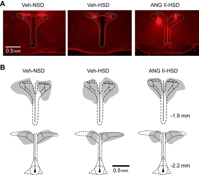
Fig. 7.
Histological verification of paraventricular nucleus microinjection sites. A: representative photomicrographs of histological sections through the PVN showing the location and distribution of fluorescent (rhodamine) nanospheres coinjected with muscimol. B: schematic drawings of microinjection sites targeting the PVN in rats (n = 6/group) infused with vehicle (Veh) and consumed a normal-salt diet (NSD; left) or high-salt diet (HSD; middle) or infused with ANG II and consumed a HSD (right). Gray regions indicate the overlapping distributions of PVN-injected nanospheres within each group. Nanosphere distribution was similar across groups. Stereotaxic coordinates on the right are with respect to bregma.

