Fig. 3.
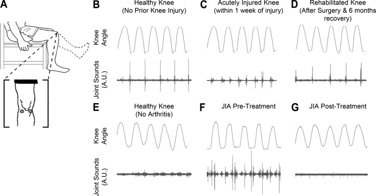
Seated unloaded knee flexion/extension acoustical emission data in two cases: knee injury rehabilitation (B–D) and juvenile idiopathic arthritis (JIA; E–F). A: locations (lateral and medial sides of the patella) of the microphones, electret and piezoelectric for B–D and E–F, respectively. B: sex and age matched control’s (with no prior knee injuries) acoustic patterns. A repetitive click is seen at a similar joint angle throughout the range of motion. C: within a week of an operable ACL/meniscus tear we notice less consistent high frequency peaks. D: after surgery to repair the injury and 6 mo of rehabilitation, we see a return of an acoustic pattern like the healthy subject in B. E: sex and aged-matched control to the patient with JIA. F: this patient with JIA (13-yr-old girl) initially presented to the clinic with poorly controlled arthritis in her knee, and acoustic emissions from the knee at the time of presentation were recorded. G: patient with JIA after undergoing treatment with methotrexate and injected corticosteroids on a return visit. The acoustic profile appears to have reverted more closely to that of the healthy control in E. This improved acoustic pattern mirrors an improved clinical picture as described by the treating rheumatologist.

