
Fig. 2.
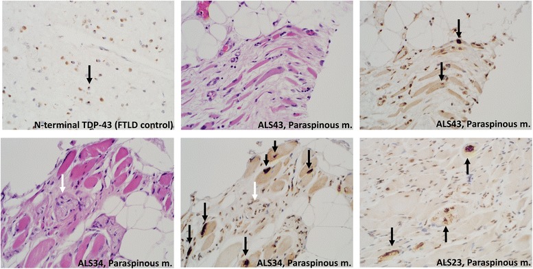
N-terminal TDP-43 immunohistochemistry in a control brain (frontotemporal lobar degeneration) and three ALS muscle samples shown to have pTDP-43-reactive inclusions. N-terminal TDP-43 immunohistochemistry reveals cytoplasmic inclusions (black arrows), as demonstrated separately with pTDP-43 immunohistochemistry. There is a loss of normal nuclear staining in affected myofibers. In sample ALS34 (bottom left) a small nerve is present (white arrow), which does not show pathologic staining in the adjacent panel (white arrow). All images are photographed at 400×