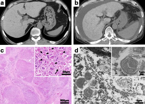
Fig. 1.

a Computed tomographic scan shows diffuse high attenuation of the liver parenchyma (96 Hounsfield units). b Massive ascites and splenomegaly were found, in addition to diffuse high attenuation of the liver parenchyma. c Regenerative nodules and well-developed bridging fibrosis were observed (hematoxylin and eosin stain, magnification × 20), as were marked neutrophilic infiltration, a remarkable amount of Mallory bodies (arrowheads), and ballooned hepatocytes (arrow) (hematoxylin and eosin stain, magnification × 200). d Numerous whorled or lamellar inclusions in lysosomes were detected by electron microscopy
