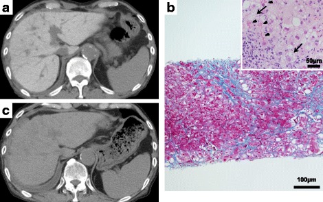
Fig. 2.

a Computed tomogram shows diffuse high attenuation of the liver parenchyma (120 Hounsfield units). b Distinct collagen deposition is seen in the periportal, perivenular, and pericellular locations, which formed bridging fibrosis (Azan stain, magnification × 100). There were numerous Mallory bodies (arrowheads) as well as hepatocellular ballooning (arrows) and mild macrovesicular and microvesicular fatty changes. Mild lymphocytic and neutrophilic infiltration was also observed (hematoxylin and eosin stain, magnification × 400). c After discontinuation of amiodarone, the patient’s liver density dramatically improved to a normal level (45 Hounsfield units) during the course of 9 months
