Fig. 3.
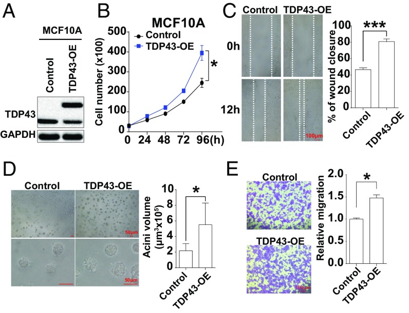
Overexpression of TDP43 promotes mammary epithelial cell proliferation and malignancy. (A) TDP43 protein levels in MCF10A cells expressing Flag-TDP43 or vehicle control by Western blotting. (B) Cell growth increased upon TDP43 overexpression in the cell-growth curve analysis. (C) Wound-healing assay in MCF10A cells stably overexpressing Flag-TDP43 or vehicle control. (D) 3D cell culture of MCF10A cells overexpressing Flag-TDP43 or vehicle control for 10 d. (E) Transwell migration assays in MCF10A cells expressing Flag-TDP43 or vehicle vectors. Data are shown as averages ± SEM for three independent measurements. *P < 0.05, ***P < 0.001.

