Fig. 7.
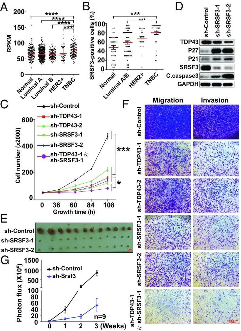
Knockdown of SRSF3 inhibits TNBC progression. (A) Transcript expression levels of SRSF3 in normal tissue and different breast cancer subtypes in the TCGA. (B) Quantification of the percentage of SRSF3-positive staining in breast cancer subtypes by immunohistochemical staining in tissue chips. (C) Cell-growth inhibition upon TDP43, SRSF3, or TDP43 plus SRSF3 knockdown by shRNAs. (D) Western blotting of cell-cycle and apoptosis-related proteins. (E) Tumor size after 2 mo of mammary fat pad growth in the xenograft assay. Each experimental group contained six mice. (F) Cell migration and invasion levels by Transwell assay. (G) Statistical analysis of fluorescence density of metastasis ability following tail-veil injection of 4T1 cells expressing luciferase (n = 9). Data are shown as averages ± SEM for three independent measurements, except in A. ***P < 0.001. All data shown represent MDA-MB231 cells, except where labeled.

