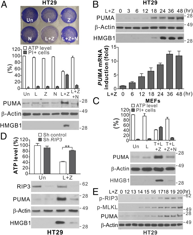
Fig. 1.
PUMA is induced in RIP1/RIP3-dependent necroptosis. (A) HT29 colon cancer cells were treated with the control DMSO (Un), LBW242 (L; 2 μM), z-VAD (Z; 10 μM), Necrostatin-1 (N; 20 μM), or the indicated combinations. (Upper) Crystal violet staining at 48 h. (Middle) ATP levels and PI staining at 48 h. (Lower) Western blots of PUMA in cell lysates and HMGB1 in 20 μL of cell culture medium at 24 h. (B) HT29 cells were treated with LBW242 and z-VAD (L+Z) as in A. (Upper) PUMA expression and HMGB1 release. (Lower) PUMA mRNA expression. (C) MEFs were treated with LBW242 (L; 2 μM), alone or in combination with TNF-α (T; 20 ng/mL), z-VAD (Z; 10 μM), or Necrostatin-1 (N; 20 μM). ATP levels and PI staining (Upper) and PUMA expression and HMGB1 release (Lower) were analyzed as in A. (D) HT29 cells stably expressing control or RIP3 shRNA were treated and analyzed as in A. (E) Western blots of phospho-RIP3 (p-RIP3; S227), phospho-MLKL (p-MLKL; S358), and PUMA in HT29 cells treated with L+Z at the indicated time points. Values in A–D are expressed as mean ± SD. n = 3. **P < 0.01.

